Cardiac Investigations
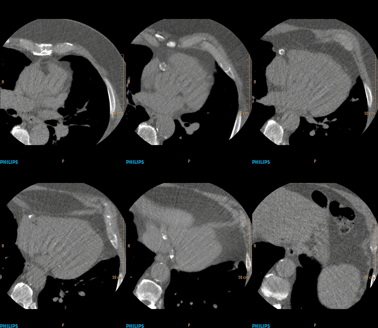
A coronary artery calcium (CAC) scan
Coronary Calcium Scan
A CT Coronary Calcium test uses x-rays to obtain images of the heart to identify “hardening” of the coronary arteries. Calcium appears in the heart’s blood vessels as a marker of plaque that has been present for many years.
This is a relatively fast test and does not require any injections. You will be asked to lie flat on the CT table and some ECG leads are attached using sticky patches on your skin. This allows the scanner to be synchronised with the hear rhythm The table then goes into the “donut” of the CT machine. The CT scanner does deliver a small dose of radiation that approximately the same as a mammogram. The images are presented as slices taken through your body which allows the doctors to assess the calcium in the arteries. This can counted and quantified to provide a score, which is useful in estimating the chance of a heart attack in the future.
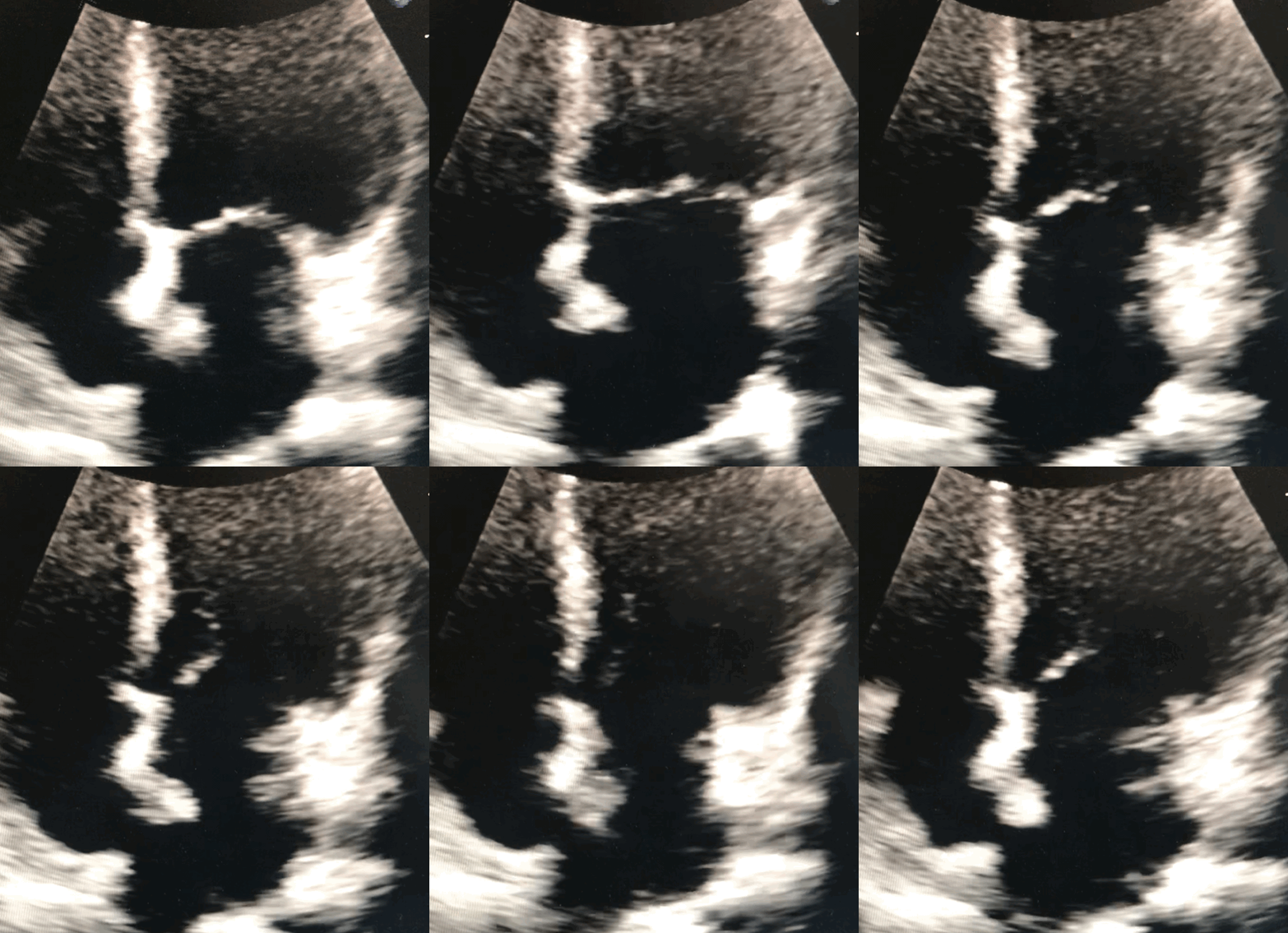
Echocardiography
Also known as echo, or trans-thoracic echo, an echocardiogram uses ultrasound waves to image the heart. The test takes around 40 minutes requires an ultrasound probe and gel to be placed on several locations around your chest. This test provides information on heart size and function. It allows assessment of the valves and flows within the heart.
Cardiac MRI
Magnetic resonance imaging obtains images using a strong magnetic field. There is no radiation or x-rays involved. This is the best test for assessing the function and composition of the heart muscle. The test can take up to 90 minutes.


